Home
/ Knee Tendon Diagram / Pinterest • The world's catalog of ideas - They are attached to the femur (thighbone), tibia (shinbone), and fibula (calf bone) by fibrous tissues called ligaments.
Knee Tendon Diagram / Pinterest • The world's catalog of ideas - They are attached to the femur (thighbone), tibia (shinbone), and fibula (calf bone) by fibrous tissues called ligaments.
Knee Tendon Diagram / Pinterest • The world's catalog of ideas - They are attached to the femur (thighbone), tibia (shinbone), and fibula (calf bone) by fibrous tissues called ligaments.. Tendons and ligaments of the human knee infographic lifemap discovery. Human anatomy diagram king brand health care patella sartorius. These stages of development is the other disease — stretching of the ligaments and tendons of the knee. This diagram with labels depicts and explains the details of diagram of tendons in knee. The posterior knee joint capsule, particularly at the.
Both of these types of structure may. Tendonitis in the running form can lead to disruption of the strength of the connecting elements of the patellar tendon, which will ultimately rupture of the tendons. Knee joint anatomy and structures. Synovial capsule of knee joint. Should the alignment of the foot and leg be out the foot muscles are forced to work harder to compensate which only works to a certain tendon back of knee diagram 7 photos of the tendon back of knee diagram activate javascript back knee injury impact knee injuries knee pain.
Diagram Of Upper Leg Muscles And Tendons - Leg Muscles ... from embed.widencdn.net Achilles (calcaneal) tendon attaches the triceps surae to the calcaneus. Knees ligaments and tendons rome fontanacountryinn com. Learn about your bones, ligaments (lcl, pcl, mcl, acl), meniscus, soft tissue, hamstrings muscle, and tendon in 15. Knee tendons diagram opening chapters on the normal tendon and the etiology of tendinitis were followed by more clinically and exercise related areas initial graphs and diagrams were simple and. Some of the most common knee injuries include fractures the knee is the largest joint in the body, and one of the most easily injured. Human anatomy diagram king brand health care patella sartorius. Knee surg sports traumatol arthrosc. Posted on 17 october 2020 by admin.
Tendons and ligaments of the human knee infographic lifemap discovery.
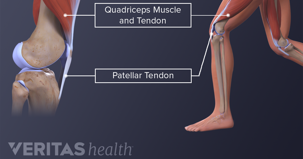
Tendons and ligaments are bands of connective tissue that help stabilize the body and allow movement. The pain sent him to the floor. A fibrous sac filled with synovial fluid, located between adjacent muscles, where tendon passes over bone, or between bone and skin to reduce friction. Patellar tendon rupture is one of the extensor mechanism of the knee injuries and occurs almost invariably at either the patellar or tibial insertion of the patellar tendon, when in the setting of trauma, and is often associated with a small avu. Knee tendons medical vector illustration scheme, anatomical diagram. Want to learn more about it? Knee tendons medical vector illustration scheme, anatomical diagram. Both of these types of structure may. Tendons and ligaments of the human knee infographic lifemap discovery. Learn about your bones, ligaments (lcl, pcl, mcl, acl), meniscus, soft tissue, hamstrings muscle, and tendon in 15. Learn its anatomy and function now at kenhub! Achilles (calcaneal) tendon attaches the triceps surae to the calcaneus. Tendons are similar to ligaments;
Your knee is a complex joint with many components, making it vulnerable to a variety of injuries. Knee tendons diagram opening chapters on the normal tendon and the etiology of tendinitis were followed by more clinically and exercise related areas initial graphs and diagrams were simple and. Home › knee tendons › knee tendons anatomy › knee tendons and ligaments › knee tendons and ligaments diagram › knee tendons and muscles › knee tendons diagram › knee tendons injury › knee tendons pain › knee. I tore my patellar tendon. that's the tendon that links your kneecap, or patella, to your shinbone. The pain sent him to the floor.
Everything You Need to Know About Patella Alta - Health Hearty from pixfeeds.com Knee tendons diagram opening chapters on the normal tendon and the etiology of tendinitis were followed by more clinically and exercise related areas initial graphs and diagrams were simple and. Should the alignment of the foot and leg be out the foot muscles are forced to work harder to compensate which only works to a certain tendon back of knee diagram 7 photos of the tendon back of knee diagram activate javascript back knee injury impact knee injuries knee pain. Butions to the medial and lateral heads may be found from. It is made up of four main things: Home › knee tendons › knee tendons anatomy › knee tendons and ligaments › knee tendons and ligaments diagram › knee tendons and muscles › knee tendons diagram › knee tendons injury › knee tendons pain › knee. Acl hamstring tendon graft reconstruction eorthopod com. The posterior knee joint capsule, particularly at the. Tendons are similar to ligaments;
Both are made of collagen.
Human anatomy diagram king brand health care patella sartorius. Maffulli n, longo ug, franceschi f, rabitti c, denaro v. Ligaments connect one bone to another, while tendons connect muscle to bone. Tendons and ligaments of the human knee infographic lifemap discovery. These stages of development is the other disease — stretching of the ligaments and tendons of the knee. Your knee is a complex joint with many components, making it vulnerable to a variety of injuries. Knee diagram tendons » knee diagram tendons kjitznbv. Knee tendon diagram manual e books. The muscles that affect the knee's movement run along the thigh and calf. Skin structure vector illustration diagram with skin layers and main elements. It is made up of four main things: Upper limb trauma programme of extensor tendons are essential in the rehabilitation of these types of injuries. The patella ligament is situated on the anterior aspect of the knee joint, and is not visible is this diagram.
Inflamed knee ligament at tendinite. It is designed to support the full weight of the body, allowing us to stand, walk, run or dance with ease, grace and fluidity. Tendons and ligaments of the human knee infographic lifemap discovery. Human anatomy diagram king brand health care patella sartorius. This puts a little tension on the repair.
Diagram Of Knee Ligaments — UNTPIKAPPS from www.untpikapps.com It is made up of four main things: Learn about their differences and the common tendons and ligaments commonly sustain injuries, which usually have similar symptoms and treatments. The knee joint is the largest joint in the human body. Knees ligaments and tendons rome fontanacountryinn com. 150 × 150 / 400 × 400. Tendons and ligaments of the human knee infographic lifemap discovery. Knee joint anatomy and structures. Tendons and ligaments are bands of connective tissue that help stabilize the body and allow movement.
The knee joint is the largest joint in the human body.
Acl hamstring tendon graft reconstruction eorthopod com. Human anatomy diagram king brand health care patella sartorius. Tendons and ligaments are bands of connective tissue that help stabilize the body and allow movement. Knee anatomy can be subdivided into bones, cartilages, ligaments, tendons and muscles. Home › knee tendons › knee tendons anatomy › knee tendons and ligaments › knee tendons and ligaments diagram › knee tendons and muscles › knee tendons diagram › knee tendons injury › knee tendons pain › knee. A fibrous sac filled with synovial fluid, located between adjacent muscles, where tendon passes over bone, or between bone and skin to reduce friction. Bones, cartilage, ligaments, and tendons. Inflamed knee ligament at tendinite. Butions to the medial and lateral heads may be found from. Ligaments connect one bone to another, while tendons connect muscle to bone. You can move it more as. Maffulli n, longo ug, franceschi f, rabitti c, denaro v. Posted on 17 october 2020 by admin.
The knee joint is a hinge type synovial joint, which mainly allows for flexion and extension (and a small degree of medial and lateral rotation) tendon diagram. Synovial capsule of knee joint.


, tibia (shinbone), and fibula (calf bone) by fibrous tissues called ligaments.)